Introduction
Once a ligand from one cell has bound to a receptor on another cell, is the signaling process complete?
If we’re talking about intracellular receptors, which bind their ligand inside of the cell and directly activate genes, the answer may be yes. In most cases, though, the answer is no—not by a long shot! For receptors located on the cell membrane, the signal must be passed on through other molecules in the cell, in a sort of cellular game of “telephone.”
The chains of molecules that relay signals inside a cell are known as intracellular signal transduction pathways. Here, we’ll look at the general characteristics of intracellular signal transduction pathways, as well as some relay mechanisms commonly used in these pathways.
Binding Initiates a Signaling Pathway
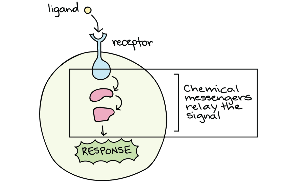
When a ligand binds to a cell-surface receptor, the receptor’s intracellular domain (part inside the cell) changes in some way. Generally, it takes on a new shape, which may make it active as an enzyme or let it bind other molecules.
The change in the receptor sets off a series of signaling events. For instance, the receptor may turn on another signaling molecule inside of the cell, which in turn activates its own target. This chain reaction can eventually lead to a change in the cell’s behavior or characteristics, as shown in the cartoon below.
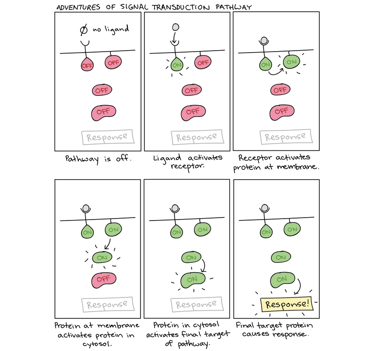
Because of the directional flow of information, the term upstream is often used to describe molecules and events that come earlier in the relay chain, while downstream may be used to describe those that come later (relative to a particular molecule of interest). For instance, in the diagram, the receptor is downstream of the ligand but upstream of the the proteins in the cytosol. Many signal transduction pathways amplify the initial signal, so that one molecule of ligand can lead to the activation of many molecules of a downstream target.
Phosphorylation Example: MAPK Signaling Cascade
To get a better sense of how phosphorylation works, let’s examine a real-life example of a signaling pathway that uses this technique: growth factor signaling. Specifically, we’ll look at part of the epidermal growth factor (EGF) pathway that acts through a series of kinases to produce a cellular response.
This diagram shows part of the epidermal growth factor signaling pathway:
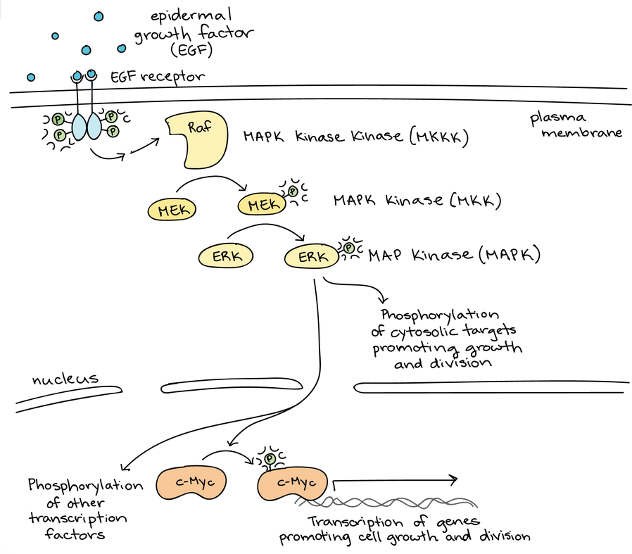
Phosphorylation (marked as a P) is important at many stages of this pathway.
Phosphorylation
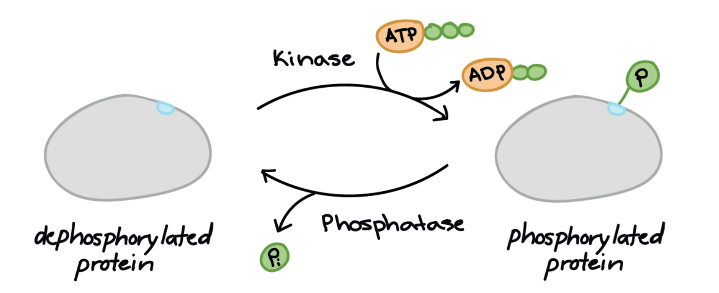
The cartoon above features a bunch of blobs (signaling molecules) labeled as “on” or “off.” What does it actually mean for a blob to be on or off? Proteins can be activated or inactivated in a variety of ways. However, one of the most common tricks for altering protein activity is the addition of a phosphate group to one or more sites on the protein, a process called phosphorylation.
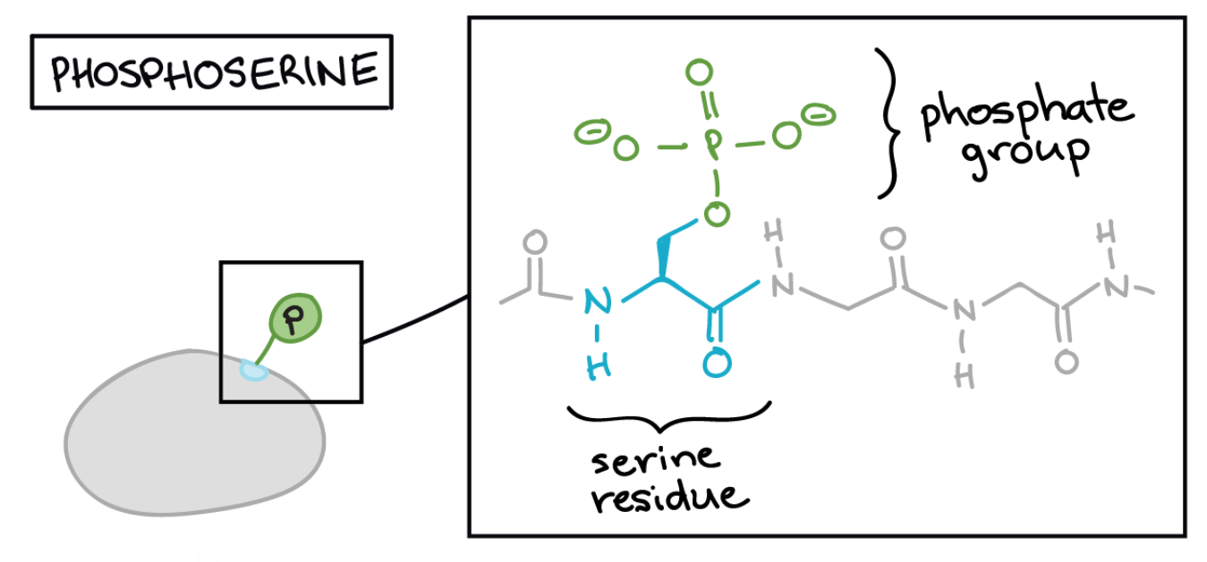
Phosphate groups can’t be attached to just any part of a protein. Instead, they are typically linked to one of the three amino acids that have hydroxyl (-OH) groups in their side chains: tyrosine, threonine, and serine. The transfer of the phosphate group is catalyzed by an enzyme called a kinase, and cells contain many different kinases that phosphorylate different targets.
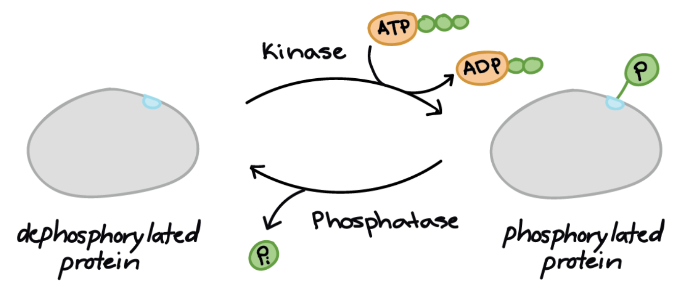
Phosphorylation often acts as a switch, but its effects vary among proteins. Sometimes, phosphorylation will make a protein more active (for instance, increasing catalysis or letting it bind to a partner). In other cases, phosphorylation may inactivate the protein or cause it to be broken down.
In general, phosphorylation isn’t permanent. To flip proteins back into their non-phosphorylated state, cells have enzymes called phosphatases, which remove a phosphate group from their targets.
Phosphorylation Example: MAPK Signaling Cascade
To get a better sense of how phosphorylation works, let’s examine a real-life example of a signaling pathway that uses this technique: growth factor signaling. Specifically, we’ll look at part of the epidermal growth factor (EGF) pathway that acts through a series of kinases to produce a cellular response.
This diagram shows part of the epidermal growth factor signaling pathway:
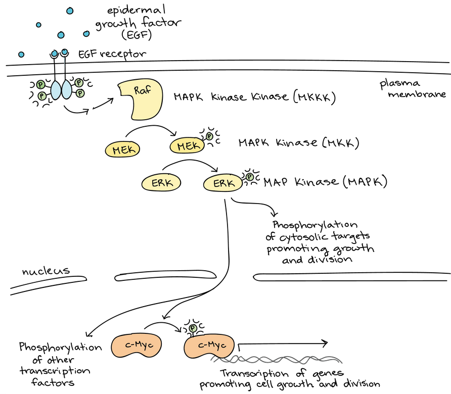 Phosphorylation (marked as a P) is important at many stages of this pathway.
- When growth factor ligands bind to their receptors, the receptors pair up and act as kinases, attaching phosphate groups to one another’s intracellular tails.
- The activated receptors trigger a series of events (skipped here because they don’t involve phosphorylation). These events activate the kinase Raf.
- Active Raf phosphorylates and activates MEK, which phosphorylates and activates the ERKs.
- The ERKs phosphorylate and activate a variety of target molecules. These include transcription factors, like c-Myc, as well as cytoplasmic targets. The activated targets promote cell growth and division.
Together, Raf, MEK, and the ERKs make up a three-tiered kinase signaling pathway called a mitogen-activated protein kinase (MAPK) cascade. (A mitogen is a signal that causes cells to undergo mitosis, or divide.) Because they play a central role in promoting cell division, the genes encoding the growth factor receptor, Raf, and c-Myc are all proto-oncogenes, meaning that overactive forms of these proteins are associated with cancer.
MAP kinase signaling pathways are widespread in biology: they are found in a wide range of organisms, from humans to yeast to plants. The similarity of MAPK cascades in diverse organisms suggests that this pathway emerged early in the evolutionary history of life and was already present in a common ancestor of modern-day animals, plants, and fungi.
Second Messengers
Although proteins are important in signal transduction pathways, other types of molecules can participate as well. Many pathways involve second messengers, small, non-protein molecules that pass along a signal initiated by the binding of a ligand (the “first messenger”) to its receptor.
Second messengers include ions; cyclic AMP (cAMP), a derivative of ATP; and inositol phosphates, which are made from phospholipids.
Calcium Ions
Calcium ions are a widely used type of second messenger. In most cells, the concentration of calcium ions () in the cytosol is very low, as ion pumps in the plasma membrane continually work to remove it. For signaling purposes, may be stored in compartments such as the endoplasmic reticulum.
In pathways that use calcium ions as a second messenger, upstream signaling events release a ligand that binds to and opens ligand-gated calcium ion channels. These channels open and allow the higher levels of that are present outside the cell (or in intracellular storage compartments) to flow into the cytoplasm, raising the concentration of cytoplasmic .
How does the released help pass along the signal? Some proteins in the cell have binding sites for ions, and the released ions attach to these proteins and change their shape (and thus, their activity). The proteins present and the response produced are different in different types of cells. For instance, signaling in the β-cells of the pancreas leads to the release of insulin, while signaling in muscle cells leads to muscle contraction.
Cyclic AMP (cAMP)
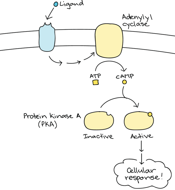
Another second messenger used in many different cell types is cyclic adenosine monophosphate (cyclic AMP or cAMP), a small molecule made from ATP. In response to signals, an enzyme called adenylyl cyclase converts ATP into cAMP, removing two phosphates and linking the remaining phosphate to the sugar in a ring shape.
Once generated, cAMP can activate an enzyme called protein kinase A (PKA), enabling it to phosphorylate its targets and pass along the signal. Protein kinase A is found in a variety of types of cells, and it has different target proteins in each. This allows the same cAMP second messenger to produce different responses in different contexts.
cAMP signaling is turned off by enzymes called phosphodiesterases, which break the ring of cAMP and turn it into adenosine monophosphate (AMP).
Inositol Phosphates
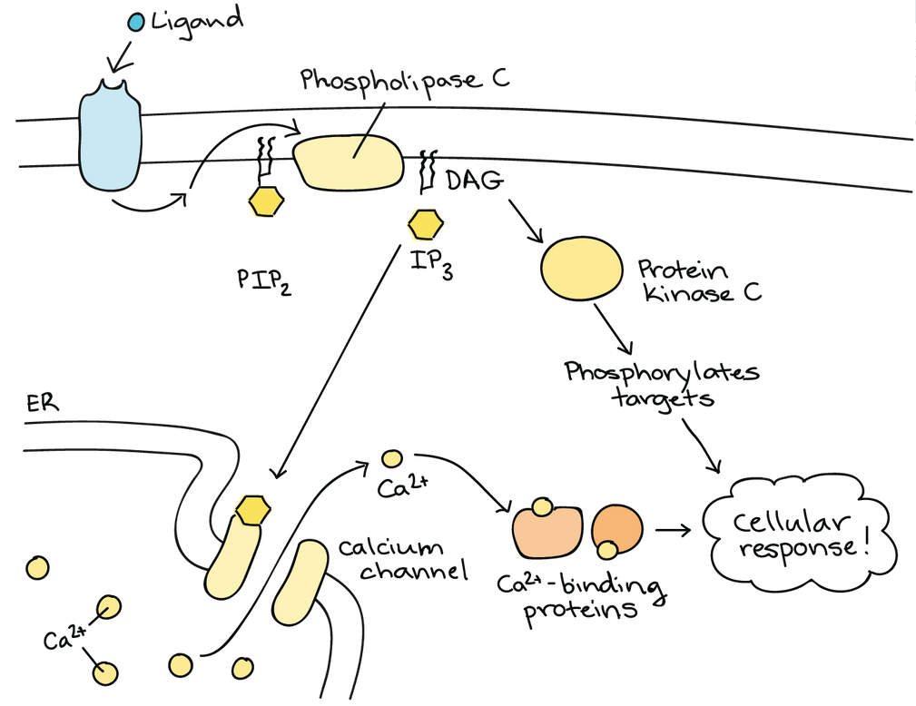
Although we usually think of plasma membrane phospholipids as structural components of the cell, they can also be important participants in signaling. Phospholipids called phosphatidylinositols can be phosphorylated and snipped in half, releasing two fragments that both act as second messengers.
One lipid in this group that’s particularly important in signaling is called . In response to a signal, an enzyme called phospholipase C cleaves (chops) into two fragments, DAG and . These fragments made can both act as second messengers.
DAG stays in the plasma membrane and can activate a target called protein kinase C (PKC), allowing it to phosphorylate its own targets. diffuses into the cytoplasm and can bind to ligand-gated calcium channels in the endoplasmic reticulum, releasing that continues the signal cascade.
And…it’s even more complicated than that!
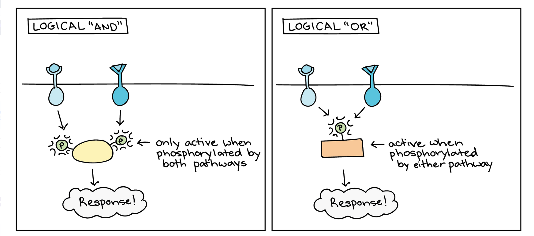
Complexity arises because pathways can, and often do, interact with other pathways. When pathways interact, they basically allow the cell to perform logic operations and “calculate” the best response to multiple sources of information. For instance, signals from two different pathways may be needed to activate a response, which is like a logical “AND.” Alternatively, either of two pathways may trigger the same response, which is like a logical “OR.”Left diagram: logical “AND” in a cell signaling pathway. An intermediate must be phosphorylated on two different residues, one targeted by each of two pathways, in order to become active and produce a response. The response only occurs if the first pathway AND the second pathway are active. Right diagram: logical “OR” in a cell signaling pathway. An intermediate must phosphorylated on a single residue in order to become active and produce a response, and either of two pathways can phosphorylate the same residue. The response occurs if the first pathway OR the second pathway is active.
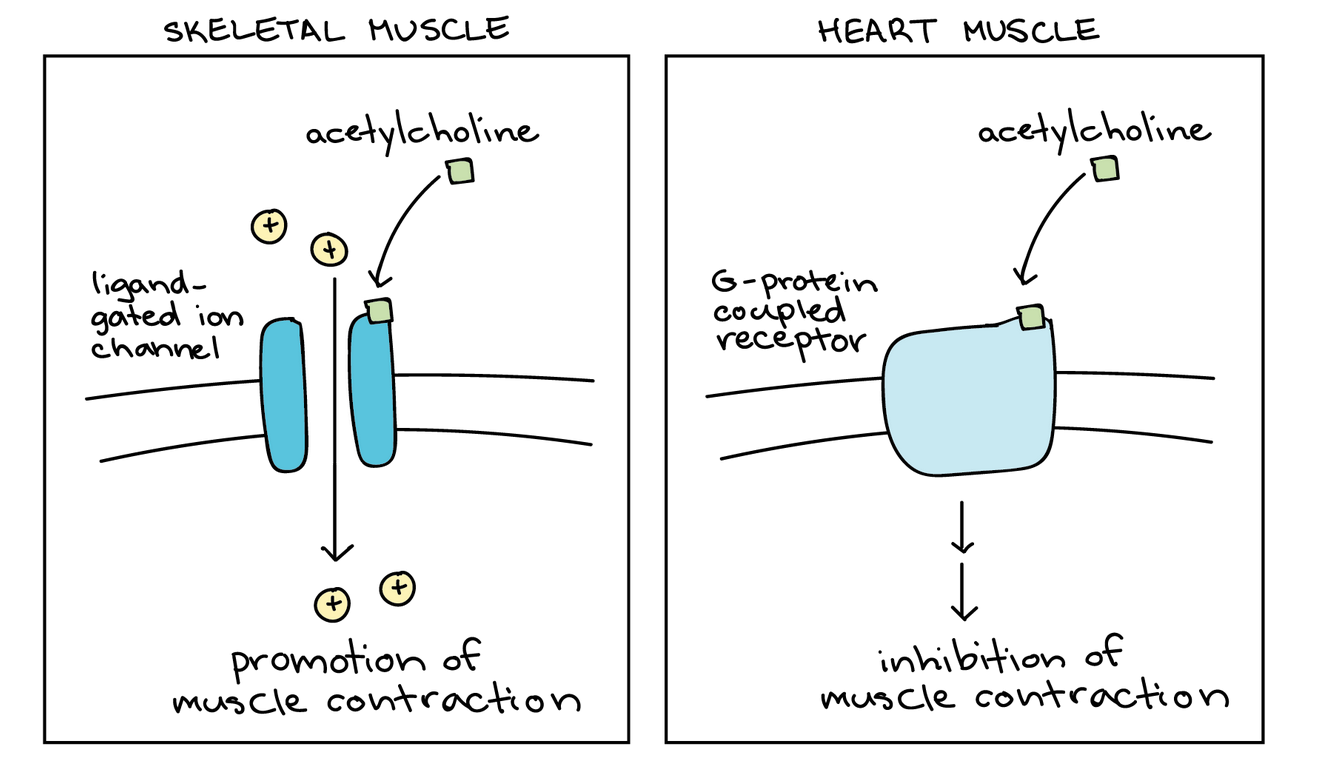
Another source of complexity in signaling is that the same signaling molecule may produce different results depending on what molecules are already present in the cell. For example, the ligand acetylcholine causes opposite effects in skeletal and heart muscle because these cell types produce different kinds of acetylcholine receptors that trigger different pathways.
These are just a few examples of the complexities that make signaling pathways challenging, but also fascinating, to study. Cell-cell signaling pathways, especially the epidermal growth factor pathway we saw earlier, are a focus of study for researchers developing new drugs against cance.